Welcome to my blog, where I summarise primary literature and provide my insights. If you’re wondering, this is not my first blog. I also had a blog during 2016 until approximately 2019, though I started it before I even got into university. There will be a big difference between my old and new blog; if you wish to compare, then here is my first blog: Superfruit Science.
We’re going to get right into it with this first post by looking at nucleotides and nucleotide metabolism and their involvement in cancer. This is FASCINATING work. Very new and innovative.
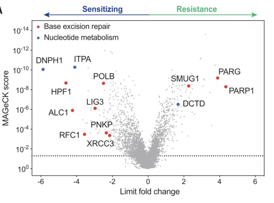
IMAGE: Figure 1A from Fugger et al.’s CRISPR-Cas9 screen showing the different genes that either have a sensitising or resistance role upon PARPi treatment (Fugger et al., 2021).
DNA Damage and Repair
DNA damage occurs frequently as cells undergo the cell cycle. There are two main sources of DNA damage: endogenous and exogenous sources. An endogenous source is when DNA replication machinery make errors as they replicate the genome. Imagine a faulty car assembly line trying to assemble a car – the end product won’t be functioning if the machines aren’t working! Exogenous sources of damage entail many carcinogens, such as smoking, asbestos, and industrial pollution. If you’re wondering how bad smoking cigarettes are, the human body is exposed to at least 60 carcinogens (Hecht, 2003; Yamaguchi, 2019). Carcinogens are cancer-causing agents that damage the DNA molecules in the affected cells.
The DNA damage repair (DDR) pathways involve a series of mechanisms that the cell uses with different types of damage to the DNA. There are two common pathways: base excision repair (BER) and nucleotide excision repair (NER). There are plenty of other methods of DNA repair as well, including mismatch repair (MMR), homologous recombination (HR), and non-homologous end-joining (NHEJ).
BRCA-mutated cancers and the PARPi problem.
The BRCA1 and BRCA2 genes are very important drivers in cancer; these two genes are essential in the DNA damage repair (DDR) pathways, thus mutations of these genes are highly associated with cancer. BRCA1 and BRCA2 are needed for repairing double-stranded DNA breaks (clean breaks in the DNA) via homologous recombination.
To put this into perspective, a study found that there was an average 65% risk for breast cancer by the age of 70 for women with BRCA1 mutations and an average of 45% risk for breast cancer by the age of 70 for women with BRCA2 mutations (Antoniou et al., 2003).
A class of medicines called poly(ADP-ribose) polymerase (PARP) inhibitors are often used to treat breast cancer. These medicines cause PARP trapping onto genomic DNA. PARP1 and PARG are involved in DNA repair, so the inhibitors prevent this by essentially trapping PARP1 into the DNA in order to stop it from repairing the cancer cell. This class of chemotherapy has definitely helped overall, but there is still an overwhelming amount of patients in which the cancer resists medication and continues growing.
CRISPR Screen for PARPi sensitivity and resistance
We’ll now dive into our main study focus for this topic looking at nucleotide metabolism and how all of the above relate to nucleotides (Fugger et al., 2021).
Fugger et al. (2021) started off investigating PARPi resistance by conducting a genome-wide CRISPR-Cas9 screen. CRISPR-Cas9 genome-wide screening is an experimental method that looks at gene expression of the entire genome of the cell samples based off of the CRISPR-Cas9 system present in bacteria that we use for gene editing and screening.
The system bacteria use consist of CRISPR (which stands for clustered regularly interspaced short palindromic repeats) DNA sequences, which are sequences from bacteriophages that previously infected the bacterium. Think of this like our flu antibodies that we store in case of reinfection – thats how this works. The CRISPR sequences enable bacteria to detect and remove new DNA from bacteriophages (viruses that infect bacteria) in order to prevent re-infection and any other future infections from bacteriophages. Cas9 is an enzyme that uses CRISPR sequences to open the DNA and remove the new virus DNA. It’s like a prototype of our immune systems!
NOTE: I will be referring to a lot of different cells in this blog. The important thing is that when I add ‘-/-‘ then it means that the gene mentioned before ‘-/-‘ is therefore not expressed in that cell line for whatever reason (knocked out or depleted or no expression).
Fugger et al. (2021) conducted a genome wide screen using cells generated via CRISPR gene editing, known as HR-deficient e-HAP MUS81-/- cells. These cells were treated with a PARPi known as olaparib for 10 days, and their sensitivity/resistance was determined; the researchers looked at which genes may be involved in the sensitivity or resistance of these cells.
Resistance was observed relating to PARP1, PARG, SMUG1 and DCTD (we’ll expand on the latter two genes later on). Sensitivity, however, was observed with DNPH1 and ITPA; these two were associated with increasing sensitivity in the cells, which can be a huge advantage to re-sensitise cancer in the future if they start responding PARPi.
‘What is the relevance of this?’ you may ask. Well, DNPH1’s biological function has never been investigated, and it’s very curious to have an unknown gene impacting the sensitivity of cancer cells to chemotherapy, especially as this has a therapeutic potential. Dr Fugger’s team were intrigued and dived deep into this.
DNPH1 and ITPA – New Players On The Field?
Fugger et al. (2021) found that DNPH1 is involved with nucleotide metabolism. Nucleotides are the building blocks of DNA. Imagine a wall with bricks, you can’t have that wall protecting you unless you have all the bricks correctly assorted. DNPH1 is specifically found to be a nucleotide sanitiser, meaning that it removes available modified nucleotides, as they can be dangerous if re-inserted into DNA. This study unravelled a whole new pathway in which nucleotides are metabolised to prevent their incorporation to DNA.
DNPH1 and ITPA are associated with sensitivity to PARPi as we saw in the screen I highlighted above. The study focusses on DNPH1 (and not as much on ITPA, which I will discuss later on). When DNPH1 was depleted or knocked out in BRCA1-/- cells or BRCA2-/- cells, they showed an increased sensitivity to PARPi treatment. This was exclusive to PARPi class drugs and not other DNA-damaging agents. BRCA wild-type (no BRCA mutation) cells had no change in sensitivity. This indicated that loss of DNPH1 sensitises *only* BRCA-deficient cells to PARPi.
hmdUMP Is DNPH1’s Biological Target
Fugger et al. (2021) found that DNPH1 has a biological target upon which it acts (hmdUMP).
They found this by conducting a metabolic analysis. DNPH1-/- cells had increased hmdU, which is a cytotoxic nucleoside produced via altering (deamination – removing an amine group in chemical terms) of hmdC after epigenetic regulation or from oxidative damage to thymidine (a base in DNA). This suggested that DNPH1 acts on hmdUMP and hydrolyses it into hmdU to prevent it’s incorporation to the genome.
So if DNPH1 reduces toxic nucleosides from entering the genome, therefore a loss of DNPH1 will increase toxicity, especially with addition of PARPi treatment.
But How is PARPi Treatment Linked to The Function of DNPH1?
The Link
MUS81-/- cells were given different treatments with different modified nucleosides. Nucleosides are similar to nucleotides, except they lack a phosphate group. hmdU is a nucleoside that is converted to hmdUMP when phosphorylated and incorporated into DNA. DNPH1acts on hmdUMP, not hmdU, but the study often used hmdU. When hmdU and olaparib were given together, lethality increased. This shows that hmdU increases the efficacy of PARPi by incorporation to DNA and forming hmdUMP; a lack of DNPH1 meant that the hmdUMP was not hydrolysed. Accumulation of hmdUMP and inhibition of PARP1 (and therefore inhibition of any repairs of the damage hmdUMP induces) leads to cell death. Again, this was only in BRCA-deficient cells; BRCA WTs were still not sensitive.
When looking at DNPH1-/- cells treated with hmdC (another nucleoside; epigenetic marker), genomic hmdU was increased. Both hmdC and hmdU impacted sensitivity, so hmdU may be derived from hmdC. A picture was forming: hmdC may lead to hmdU, which leads to hmdUMP and DNA damage unless hmdUMP was hydrolysed by DNPH1 or the damage was repaired by PARP1. Adding DNPH1 to DNPH1-deficient cells reversed the hmdU-induced toxicity.
BRCA2-/- DNPH1-/- cells were sensitive to PARPi when treated with hmdU, especially at a lower dose. Less chemotherapy was needed to induce synthetic lethality.
Another gene which I mentioned previously, SMUG1, has a role in resistance. SMUG1 is involved in removing hmdU from DNA. When cells were depleted of SMUG1, there was resistance to hmdU and olaparib. BRCA2-/- DNPH1-/- cells with sensitivity to olaparib and hmdU became resistant when SMUG1 was depleted. When SMUG1 was added back, there was sensitivity restored. This indicates that both DNPH1 loss and a lack of SMUG1-dependent excision of hmdU (both lead to accumulation of hmdUMP ultimately) is the basis for synthetic lethality when PARPi treatment was given.
How does PARPi treatment work?
The basis is that PARP1 is trapped to the DNA, so it becomes associated with the chromatin, and can no longer do its job to repair DNA.
Fugger et al. (2021) confirmed this by finding an increased PARP1 chromatin association upon hmdU and PARPi treatment compared with only PARPi treatment. The action of DNPH1 increases PARP trapping. They also found increased apoptosis (Cell death) as well as activation of the DNA damage checkpoint in G2 of the cell cycle. In short, the checkpoint in which the DNA is checked for damage was found to be activated. The action of this PARP trapping occurs in SMUG1-mediated hmdU excision sites. A loss of DNPH1 and it’s action of removing these toxic nucleotides causes DNA damage, replication stress, and apoptosis. When SMUG1 was removed, all of this was reversed.
DNA replication fork collapse tops it all off
Fugger et al. (2021)’s work expanded into looking at the replication fork, which is essentially the opened DNA where active DNA replication occurs. They found that this fork collapses when hmdU and olaparib were given to DNPH1-/- cells; there is assymmetry in the fork. Replication fork collapses also lead to double-stranded breaks.
To summarise: they found that this seemingly insignificant DNPH1 enzyme actually has chaotic impacts in the DNA of cancer cells if depleted.
Inducing DNPH1 inihibition
The study wanted to see if chemical inihibition of DNPH1 could have the same impact as cell lines with DNPH1 depleted. Think of it this way: cancer cells in the patient are not able to be completely manipulated this way as easily. Cells in a plate are much easier to engineer, especially with the CRISPR gene editing that the lab used.
A competitive DNPH1 inhibitor was applied with hmdU, finding that sensitivity (and lethality) was increased in BRCA deficient cells.
Main Message
The main point of this study was that a new route investigating nucleotide metabolism actually has profound impacts on PARPi resistance. There seems to be a complex map of these enzymes and their nucleoside/nucleotide targets that are involved in PARP treatment of BRCA-deficient cancers. In particular, the study suggests that DNPH1 inhibition could be a future therapeutic advantage to exploit in order to re-sensitise resistant cancers in patients.
The final points of the study is this:
- PARPi treatments inhibit PARP1 by trapping them to DNA and preventing DNA repair in cancer.
- Cancers often have resistance to PARP inhibibtors.
- DNPH1 and ITPA were found to be involved in PARPi sensitivity, with DNPH1’s biological functional target unknown. PARP1, PARG and SMUG1 were involved in resistance.
- DNPH1’s biological functional target is hmdUMP.
- hmdC from epigenetic changes is converted to hmdU which becomes hmdUMP when incorporated into DNA.
- hmdUMP causes DNA damage
- When hmdU and PARPi was given to BRCA-deficient DNPH1-/- cells, there was increased sensitivity to the PARPi and synthetic lethality (both hmdU and PARPi were needed).
- hmdUMP accumulation due to hmdU and PARPi treatment without DNPH1 means that more DNA damage occurs than just PARPi alone or in WT cells.
- SMUG1 excises hmdU, so the depletion of SMUG1 leads to increased hmdU as well (therefore hmdUMP in BRCA-deficient DNPH1-/- cells).
- Therefore hmdU and PARPi treatment to BRCA-deficient DNPH1-/- SMUG1-depleted cells had increased sensitivity and cell death to PARPi. There was also DNA fork collapse, apoptosis, and replication stress.
- Chemical inhibition of DNPH1 in BRCA-deficient cells also had increased sensitivity
- There could be a therapeutic use for these findings, and more research needs to be done into nucleotide metabolism in cancer and even other diseases.
Epigenetic changes and oxidative damage to thymidine base -> hmdC -> hmdU-> hmdUMP -> hydrolysis by DNPH1 to prevent DNA damage
Epigenetic changes and oxidative damage to thymidine base -> hmdC -> hmdU-> hmdUMP -> DNA damage when DNPH1 is lost
My Thoughts
I have lots of ideas and thoughts and questions!
I think there is great potential here, and Fugger’s lab have made fantastic advances in this field. Before having the chance to speak to Dr Fugger myself, I had never heard of this. I think the idea that DNPH1i treatment could help resistance in patients in the future is reasonable and innovative.
Not sure if you can tell from my writing, but the study looked at a lot of different factors and did a lot of work in order to provide a story. A good research paper gives you the context and forms a story that you follow along in their data. I’m still trying to comprehend this, as it is a new concept for me. The paper is generally clear to understand, but putting together the first ideas with the progressing complex thoughts later on is still on my mind! Their data is relatively easy to interpret and it really shows the story as we go along.
However, I do have some constructive criticism/advice. They mention ITPA, but have not done much research on ITPA compared with DNPH1 as much. I would also like to see more work on chemical inhibition of DNPH1. The study doesn’t distinguish hmdUMP and hmdU and hmdCMP and hmdC very well. They first start with stating that DNPH1 acts on hmdUMP, but then they talk about hmdU. This confused me a little at first, so it could have been clearer both in text and in the figures rather than just the figures. This was a fantastic start, but theres a lot of work to do (this last sentence was less criticism and more just excitement that theres so much to do).
I have a lot of questions for the researchers and generally for all of us to think about:
- What is the relevance of using HR-deficient e-HAP MUS81-/- cells specifically? Why choose this cell line at first?
- What about genetic disorders such as Li-Fraumeni Syndrome? People with LFS in the family have cancer recurring very frequently, even in the same individuals.
- What about people who get cancer multiple times? Could there be an impact in DNPH1 and SMUG1 related sensitivity after the first cancer?
- What about liquid cancers? Same principle?
- What about different breast cancers? BRCA mutations aside, there are many different variants of cancer. What about triple negative?
- What about the G2 decatenation checkpoint? The study looked at the DNA damage checkpoint in G2, but not the decatenation checkpoint in which Topoisomerase 2a is involved in decatenating DNA.
- If hmdU/DNPH1i/PARPi were all used simultaneously in patients, what could the potential impacts be on patients? Would this impact non-cancerous cells?
- Could hmdU treatment, a cytotoxic nucleoside, induce more DNA damage in non-cancerous cells?
- In my MSc looking at the G2 decatenation checkpoint in different breast cancer and epithelial cell lines, I found that cells were not responsive to ICRF193, which is meant to inhibit Topo2a. The cells did not die at the G2 decatenation checkpoint as they should have and continued into mitosis. Could this be relevant to this study into PARPi resistance?
- In a discussion with Dr Fugger, we discussed that nucleotides can be found in diet and some gut bacteria. What impacts could this be having on the increasing prevalence of colon cancer? Could nucleotides be absorbed via the digestive system which could induce more DNA damage and increase cancer risk? Could there be different foods that increase that risk compared to other foods?
I’m not an expert in this at all, so by all means my questions could be stupid or unnecessary, but I do like to write down my ideas anyway an
References
- Antoniou, A., Pharoah, P. D., Narod, S., Risch, H. A., Eyfjord, J. E., Hopper, J. L., Loman, N., Olsson, H., Johannsson, O., Borg, A., Pasini, B., Radice, P., Manoukian, S., Eccles, D. M., Tang, N., Olah, E., Anton-Culver, H., Warner, E., Lubinski, J., Gronwald, J., Gorski, B., Tulinius, H,. Thorlacius, S., Eerola, H., Nevanlinna, H., Syrjäkoski, K., Kallioniemi, O. P., Thompson, D., Evans, C., Peto, J., Lalloo, F., Evans, D. G., & Easton, D. F. (2003). Average risks of breast and ovarian cancer associated with BRCA1 or BRCA2 mutations detected in case Series unselected for family history: a combined analysis of 22 studies. American Journal of Human Genetics., [online] 72, 1117-1130. Available at: https://pubmed.ncbi.nlm.nih.gov/12677558/https://pubmed.ncbi.nlm.nih.gov/12677558/.
- Fugger, K., Bajrami, I., Silva Dos Santos, M., Young, S. J., Kunzelmann, S., Kelly, G., Hewitt, G., Patel, H., Goldstone, R., Carell, T., Boulton, S. J., MacRae, J., Taylor, I. A., & West, S.C. (2021). Targeting the nucleotide salvage factor DNPH1 sensitizes BRCA-deficient cells to PARP inhibitors. Science, [online] 9, 156-165. Available at: https://pubmed.ncbi.nlm.nih.gov/33833118/https://pubmed.ncbi.nlm.nih.gov/33833118/.
- Hecht, S. S. (2003). Tobacco carcinogens, their biomarkers and tobacco-induced cancer. Nature Reviews Cancer, [online] 3, 733-744. Available at: https://www.nature.com/articles/nrc1190.
- Yamaguchi, N. H. (2019). Smoking, immunity, and DNA damage. Transl Lung Cancer Res, [online] 8, S3-S6. Available at: https://www.ncbi.nlm.nih.gov/pmc/articles/PMC6546629/#:~:text=Smoking%20directly%20exposes%20the%20epithelial,bronchi%2C%20and%20lung%20epithelial%20cells.

 Biological Babble
Leave a comment